Our office-based, full-service, IAC Vascular Testing (ICAVL) accredited vascular laboratory allows accurate diagnosis of vascular problems via advanced noninvasive technology. In evaluating the veins and arteries throughout the body, we aim to use effective and timely treatment for all patients.
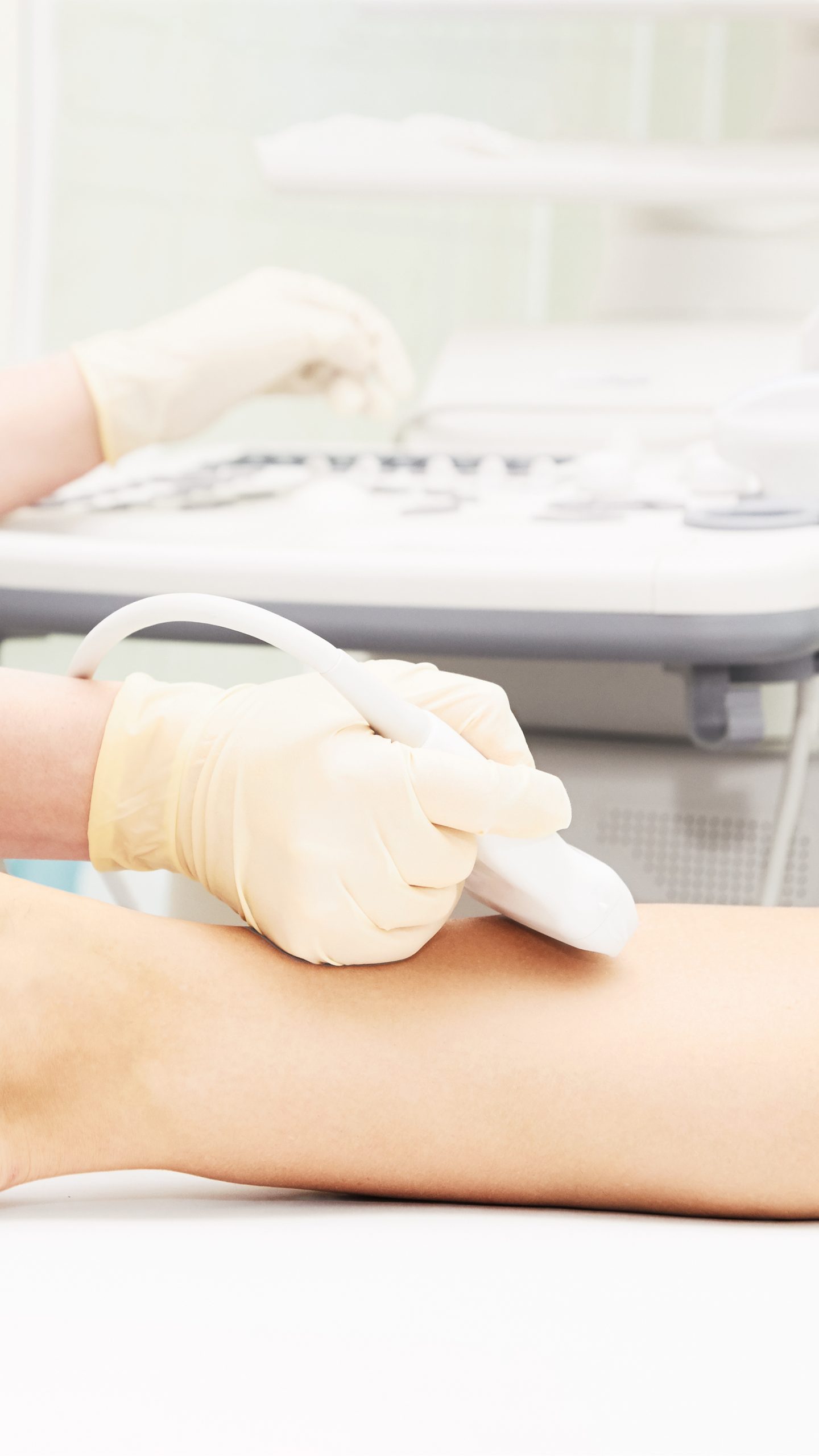
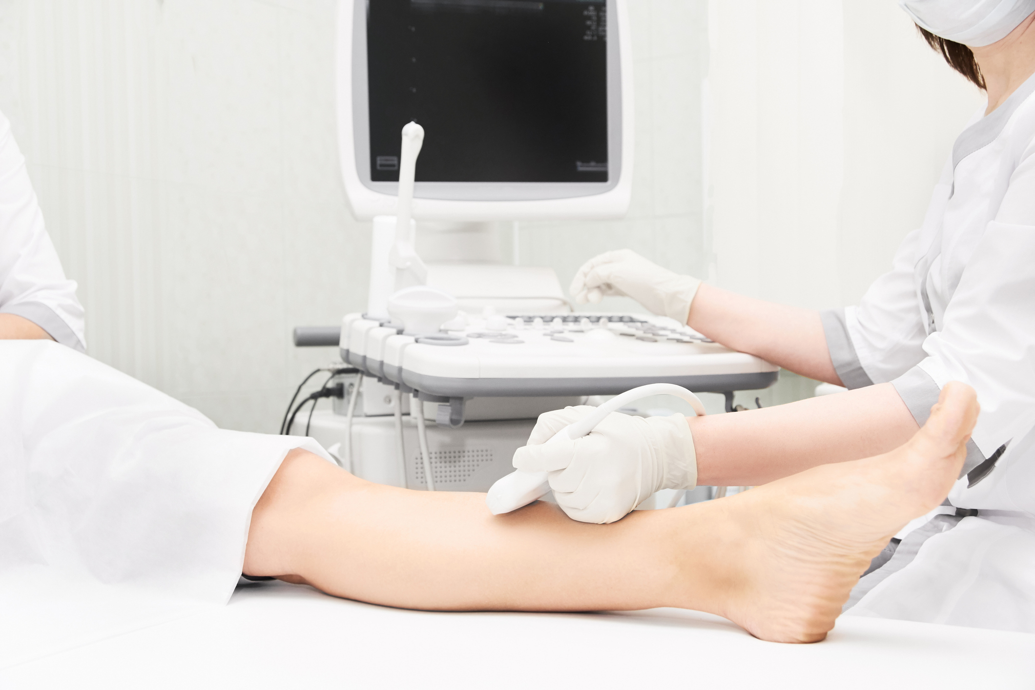
We perform several vascular lab tests in our office. They include arterial Doppler, plethysmography and ultrasound tests that help identify information about the blood vessels such as vein dysfunction, arterial blockages, aneurysms and blood clots. If necessary, our registered technologists work with your doctors to develop a treatment plan.
Vascular Services Offered Through Our Lab
We offer many services through our state-of-the-art diagnostic vascular laboratory, including:
Venous Duplex Ultrasound
A venous duplex scan captures internal images of veins returning blood to the heart. This test is painless and can identify vein dysfunction and blood clots using high-frequency sound waves through an ultrasound scan.
A lower extremity examination views the veins in the ankles and legs, while an upper extremity examination views the veins in your wrists, arms, shoulders and neck.
Arterial Duplex Ultrasound and Doppler
A duplex ultrasound combines traditional ultrasound and a Doppler ultrasound to identify blood flow, peripheral aneurysms and arterial blockages.
An arterial Doppler ultrasound records sound waves that bounce off moving objects such as blood to measure their speed and flow. Traditional ultrasound uses sound waves that create pictures by bouncing off blood vessels. Arterial Doppler studies are great for detecting abnormal flow within a blood vessel or artery.
Aortic Imaging and Duplex
Aortic imaging plays a critical role in post-surgical surveillance and treatment planning of aortic pathology. Patients experiencing aortic abnormalities require lifelong supervision, and imaging is essential in monitoring any changes or progress.
One-time duplex ultrasound screening is cost-effective and can reduce aneurysm-related mortality due to rupture. This noninvasive procedure also plays a significant role in screening patients at risk for developing abdominal aortic aneurysms (AAA).
Venous Mapping
A vein mapping examination identifies the size of the veins using ultrasound. A noninvasive test, the ultrasound uses gel, sound waves and a small probe to procure an image of the veins in the arms and legs.
This technique allows for better treatment planning and allows the doctor to see the depth, size and blood flow in these veins.
Graft Duplex
Vein grafts come from patients’ own arteries and veins located in the arm, leg or chest. A graft carries oxygen-rich blood to the heart via a new pathway.
Frequent surveillance of the vein graft is required to detect and correct the stenoses or the abnormal narrowing in a blood vessel or other tubular organ that may lead to graft failure. A new ultrasound imaging method — vein graft surveillance with tracking duplex ultrasound imaging — allows the continuous monitoring of the vein graft by displaying a panoramic view.
Our vascular surgeon, Dr. Mouhamad Annous, uses duplex ultrasounds as a noninvasive evaluation of blood flow through veins and arteries at Premier Vascular Center of Maryland. The information garnered from these tests allows us to make a diagnosis and outline a sound treatment plan.
Subclavian, Carotid and Vertebral Artery Ultrasound and Doppler Studies
A carotid/vertebral duplex test uses high-frequency ultrasound to examine blood flow within arteries such as the subclavian, vertebral and carotid arteries.
These ultrasound and Doppler studies can visualize blood vessel walls to allow surgeons and other medical professionals to devise appropriate care plans and determine the best forms of treatment.
Renal Artery Ultrasound and Duplex
A renal artery duplex uses ultrasound technology to measure blood flow. Renal artery ultrasounds examine the veins and arteries of the kidney to check for narrowing or blockages.
SMA Duplex
Superior mesenteric artery (SMA) duplex scanning is an ultrasound test that examines stomach arteries using high-frequency sound waves.

Hemodialysis Access Ultrasound
Vascular access, or hemodialysis access, is a way to reach the blood for hemodialysis. The access allows blood to travel to the dialysis machine, where it is cleaned as it traverses through a dialyzer or a special filter.
The correction of access abnormalities early may improve function and longevity of blood flow.
Evaluation and Imaging for Thoracic Outlet Syndrome
The medical team at Premier Vascular Center of Maryland is one of the few in the area to perform procedures to detect thoracic outlet syndrome (TOS).
TOS refers to a group of disorders that occur when nerves or blood vessels in the space between your first rib or thoracic outlet and your collarbone are compressed. This compression can cause numbness in the fingers and pain in your neck and shoulders.
Imaging for thoracic outlet syndrome involves using Doppler ultrasound. Your doctor may use this test to assess blood flow in the thoracic outlet or the arteries to identify any compression or blockages.
Evaluation for Raynaud’s Syndrome
Secondary Raynaud’s, also known as Raynaud’s phenomenon or Raynaud’s syndrome, is often a condition that attacks your body’s connective tissues, like rheumatoid arthritis or lupus. Primary Raynaud’s, or Raynaud’s disease, often produces mild symptoms and occurs without any other illness behind it.
Doctors use a nailfold capillaroscopy (NV) test to tell the difference between primary and secondary Raynaud’s. The doctor will look at the skin on your fingernail under a magnifier or a microscope to identify swelling or deformities of the tiny blood vessels. To look for conditions linked to secondary Raynaud’s, your doctor may order blood tests.

Schedule Your Consultation With the Premier Vascular Center of Baltimore Today
The medical team at Premier Vascular Center of Maryland is your source for high-quality vascular care.
When you chose us for vascular services, you’ll have the option for a same-day surgery after an initial consultation with Dr. Annous. He will work with you through all stages of care — consultation, surgery, and follow-up visits. The Peripheral Vascular Laboratory (PVL) at Premier Vascular Center of Maryland uses noninvasive testing, meaning there are no incisions or needle sticks for these vascular lab tests.
Reach out today to learn more about our vascular lab services and schedule your first consultation with one of our vascular surgeons.