Vascular testing often involves ultrasound, a noninvasive, versatile procedure used to examine circulation in the body’s blood vessels. Sound waves bounce off the blood cells during a vascular test as they penetrate the tissues being analyzed.
At Premier Vascular Center of Maryland, we are proud to provide advanced and accurate vascular testing with the most modern technology in Baltimore. Our vascular laboratory has maintained full accreditation through the Intersocietal Accreditation Commission (ICAVL). The vascular testing is performed by Certified Vascular Technologists.
What Does Vascular Mean?
The word vascular refers to the blood vessels which carry blood from the heart to the body’s organs and from the organs back to the heart, also known as the body’s circulatory system.
What Is Vascular Disease?
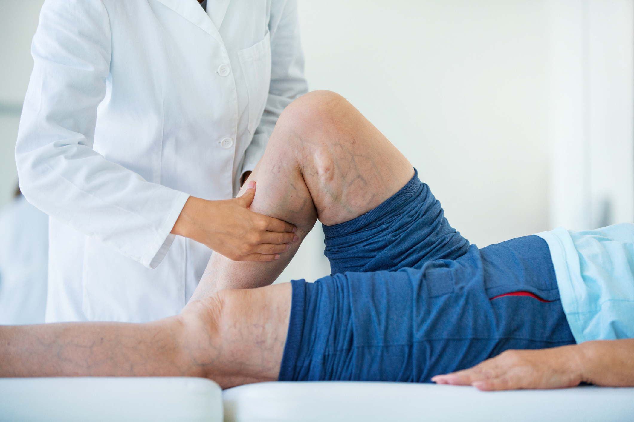
The unhealthy changes which occur in our blood vessels as we age are called vascular diseases. Examples include blood clots in the veins, varicose veins and plaque build-up in the arteries.
What Is Vascular Ultrasound?
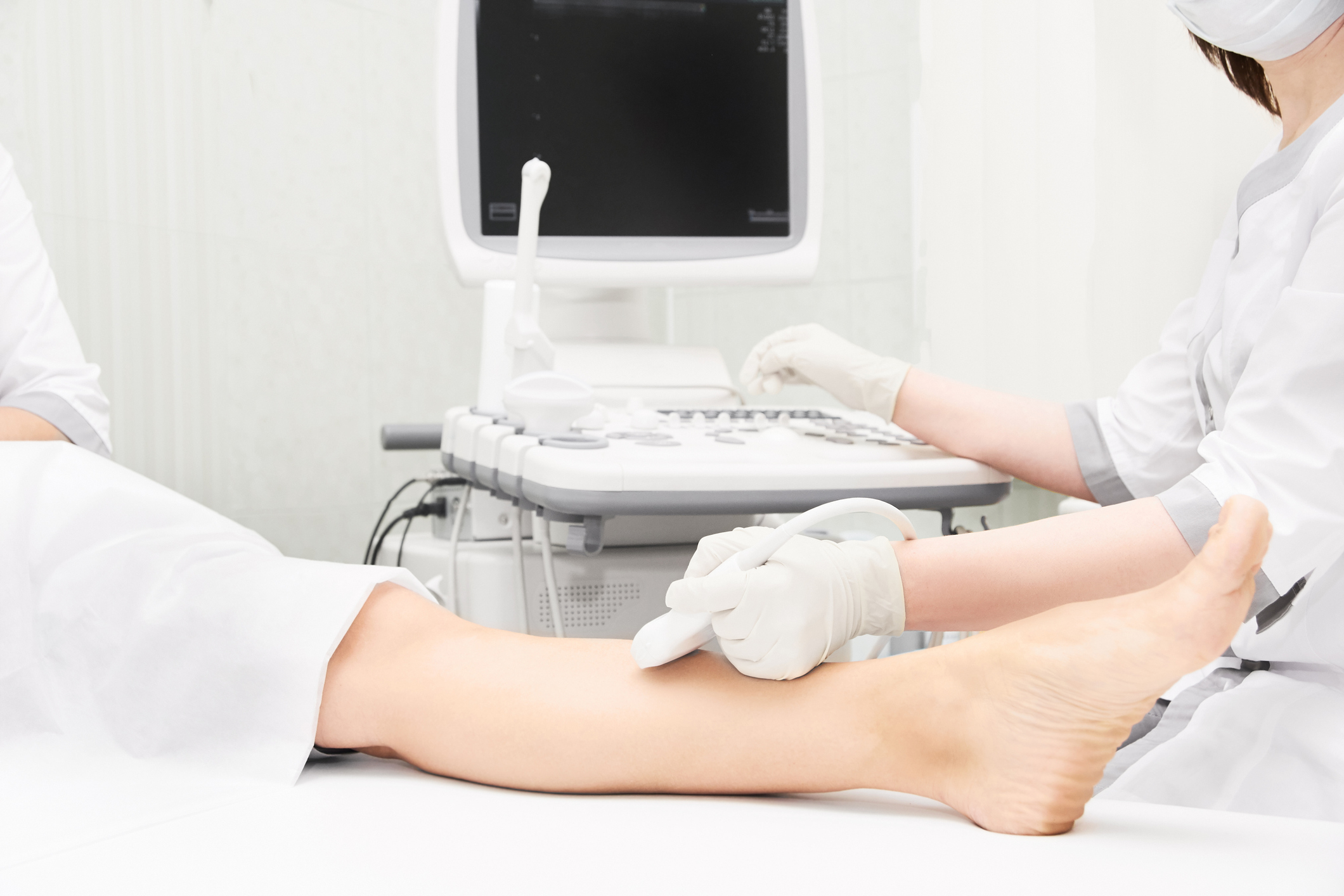
A vascular ultrasound, also known as a Doppler or duplex study, uses high-frequency sound waves to generate images of blood flow within the arteries and veins.
For example, if a vascular physician wants to see if you have a blood clot in your leg, they might order a venous Doppler examination or venous duplex. Vascular ultrasound procedures are harmless and painless. These vascular tests are noninvasive, meaning they do not require anesthesia, radiation, dyes, or needles.
The information gathered through vascular ultrasounds allows physicians to diagnose various blood vessel disease and other conditions related to vascular disease.
Noninvasive vascular testing techniques performed within accredited vascular laboratories aid in the early detection of life-threatening vascular diseases.
Types of Vascular Exams
At Premier Vascular Center of Maryland, our team of Certified Vascular Technologists perform various types of vascular tests, including the following:
Venous Duplex Ultrasound and Doppler
A venous duplex or Doppler is a test that uses ultrasound to create sounds and images of blood flow. Physicians typically request these scans on the upper and lower extremities to help detect blood clots in the veins.
In an ultrasound exam, technologists place a hand-held probe against the skin. The probe emits sound waves that bounce off moving blood and tissue, then echo back to the probe. A computer translates this echo into an image for the physician to examine and interpret.
A venous ultrasound exam aims to shed light on valuable information about possible blood clots in the extremities and other venous blood flow disorders.
Arterial Duplex Ultrasound and Doppler
Like a venous ultrasound, an arterial duplex or Doppler ultrasound creates a map of the arteries in your legs using sound waves. This scan allows your physician to identify why you may be experiencing leg pain while walking and detect any narrowing of your blood vessels.
A vascular technologist may conduct an arterial ultrasound to help diagnose many conditions, including circulation problems, blocked arteries, poorly functioning valves and blood clots.

SMA Duplex
A superior mesenteric artery (SMA) duplex utilizes high-frequency sound waves to examine the arteries in your stomach. Some of the indications for SMA artery duplex imaging include:
- Abdominal bruit.
- Persistent diarrhea.
- Suspected aneurysm.
- Evaluation following surgery.
- Significant unexplained weight loss.
- Abdominal cramping and pain associated with eating.
Renal Artery Ultrasound and Duplex
A renal artery duplex examines the arteries in and around the kidneys using high-frequency sound waves. These arteries can become blocked, resulting in kidney failure or high blood pressure.
Subclavian, Carotid and Vertebral Artery Ultrasound and Doppler Studies
A carotid or vertebral duplex test examines blood flow within the subclavian, vertebral and carotid arteries and visualizes blood vessel walls using ultrasound. Information regarding the location and degree of abnormalities is obtained noninvasively.
Bypass Graft Duplex Ultrasound
A bypass graft ultrasound is a procedure that “sees” inside your body using sound waves. The scan records and views blood flow after a surgical graft procedure that uses synthetic material to bypass diseased vessels from your arms or legs.
Venous Mapping
Venous mapping uses Doppler technology to view all of the veins in the arms or legs. A physician uses ultrasound to see the depth, size and blood flow in these veins. These veins then may be used for fistula creation for hemodialysis access or creation of an arterial bypass.
Evaluation and Imaging for Thoracic Outlet Syndrome
We are one of the few facilities in the Baltimore area that evaluate and diagnose thoracic outlet syndrome (TOS).
TOS is a condition causing weakness, tingling, numbness and pain in the arms or shoulders. This originates from neck pressure against the blood vessels and nerves that go to the arm. Depending on which structure is being compressed, TOS can be arterial, venous or neurogenic.
Vascular physicians use an arterial duplex ultrasound to assess blood flow in the arteries of the thoracic outlet, or the ring just below the collarbone formed by the top ribs.
Evaluation for Raynaud’s Syndrome
Raynaud’s syndrome is a disease that affects blood flow primarily in the toes and fingers, and sometimes the nose, knees, nipples or ears.
After a thorough evaluation, a vascular physician will draft an individualized treatment plan based on the patient’s preferences, overall health and the type and extent of the disease.
Treatment for Raynaud’s syndrome can include medical therapies, surgery or lifestyle changes.

Contact Premier Vascular Center of Baltimore to Schedule a Consultation Today
If you’re looking for vascular specialists in the Baltimore area, turn to the Premier Vascular Center of Maryland. For over 30 years, our physician has provided comprehensive vascular surgical care, vascular testing services, and wound care to treat a range of vascular issues.
If you’d like to schedule vascular testing or learn more about our services, please reach out to us.